

Left Medial
| KORBINIAN BRODMANN AND THE BRODMANN AREAS
One of the greatest achievements of modern medicine and neuroscience is imaging, the ability to depict neuronal activity, which is now called brain mapping. The German neuroanatomist and psychiatrist Korbinian Brodmann (1868–1918) was the first to map the human brain. He spent days and nights looking at wafer-thin sections of human brains under a light microscope. He wanted to divide nerve cell tissue into distinct areas according to its structure and composition and found out that, for example, the cell bodies in the gray matter are distributed differently and certain types of neurons only appear in very specific regions. In 1909 he presented a map of the cerebrum with 52 areas. He called this "cytoarchitecture".Even then he was able to assign different functions to some of these areas. Today, most Brodmann areas are assigned functions, which makes them particularly interesting for working with neurofeedback. With the help of modern procedures, these areas can also be reached with the EEG. The methods used for this use the LORETA technique, a mathematical approximation that makes it possible to measure the activity of these structures by calculating the EEG and thus use it for neurofeedback.
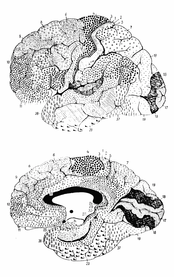
Before Brodmann's research, some areas of the brain were beginning to be identified, such as Broca's and Wernicke's areas. This came after it was found that people who had defects in these areas also suffered from speech disorders. Brodmann's mapping of the brain captures the functional areas of the cortex based on gross anatomical features and cortical microstructure. Brodmann's areas have been studied more extensively over the years and remain the best known and most used map of the cortex. Even if some of the findings from back then are no longer applicable today and have been replaced by more precise methods.
The IFEN poster about the Brodmann areas contains a QR code for each of these areas, which, after scanning, takes you to the corresponding website with the associated explanations. At the same time, you can also study the Brodmann areas without the poster. However you want to use the attractive poster. You can also use the poster as an attractive eye-catcher that adorns one or the other wall in your practice.
Download Brodmann Area Poster
| Explore brodman areas in details:
Frontal Lobe

Brodmann Area 4 — Primary Motor Cortex

Brodmann Area 6 — Premotor and Supplementary Motor Cortex

Brodmann Area 8 — Frontal Eye Fields

Brodmann Area 9 — Dorsolateral Prefrontal Cortex (superior)

Brodmann Area 10 — Frontopolar Cortex

Brodmann Area 11 — Orbitofrontal Cortex

Brodmann Area 12 — Inferior Frontal Gyrus / Ventromedial Prefrontal Cortex

Brodmann Area 44 — Pars Opercularis (Broca's Area, dominant)

Brodmann Area 45 — Pars Triangularis (Broca's Area, dominant)

Brodmann Area 46 — Dorsolateral Prefrontal Cortex (middle gyrus)

Brodmann Area 47 — Pars Orbitalis (Inferior Frontal Gyrus)
Central Region


Parietal Lobe


Temporal Lobe (lateral)


Temporal Lobe (medial)


Cingulate Cortex

Brodmann Area 23 — Posterior Cingulate Cortex (ventral)

Brodmann Area 24 — Anterior Cingulate Cortex (dorsal)

Brodmann Area 25 — Subgenual Cortex

Brodmann Area 26 — Ectosplenial Cortex

Brodmann Area 29 — Retrosplenial Region (granular)

Brodmann Area 30 — Retrosplenial Region (agranular)

Brodmann Area 31 — Dorsal Posterior Cingulate Cortex

Brodmann Area 32 — Anterior Cingulate Cortex (dorsal/rostral)

Brodmann Area 33 — Pregenual Cingulate Cortex


